From Molecules to Man
A Perspective on Size
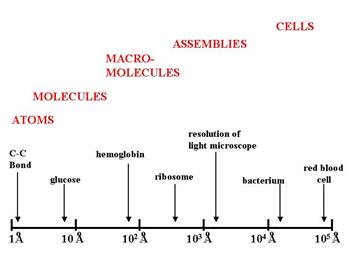
An angstrom is one ten-millionth of a millimeter, or 1×10⁻¹⁰ meters. The illustration below gives an idea of the relative scale of some of the biological structures discussed above.

The distance between two carbon atoms in a fatty acid chain is a little over one angstrom. A glucose molecule is about 9 angstroms. Bacteria are tens of thousands of angstroms. And as a rough estimate, a typical human cell might be approximately 1/100th of a millimeter which is about 1/10th the width of a human hair. For an intriguing perspective on the size of things from the smallest to the largest objects in the universe, take a look at http://htwins.net/scale2/.
Despite their microscopic size, cells have a lot going on all the time. Diagrams and photomicrographs depict cells as rigid, static sacs that are frozen in time, but if we could somehow take a trip inside a cell, we would be staggered by the beauty, complexity, and incredible activity. You can get at least a glimpse of the inner life of a cell by watching the Harvard University animation, "The Inner Life of a Cell" (full length with narration), showing leukocyte activation in inflammation. Some of the terms used in the video will be foreign to you, but the video provides a magnificent sense of the inner works of cells, and it shows cells to be dynamic structures in which many processes are taking place continually.
The Power of Polymers
One of the fundamental concepts in biology is that simple molecular structures (monomers) can be linked together to form increasingly complex structures. For example, monomers of sugars, such as glucose and fructose, can be linked together to form very large polysaccharides like starch and glycogen. Amino acids can be linked together to form polypeptides (proteins). Nucleotides can be linked together to form DNA and RNA.
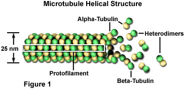
In addition to linking molecules together to form long chains, many molecules will self-assemble under appropriate conditions to form increasingly complex molecular aggregates, such as membranes or lipoproteins. And biological membranes can provide the structure for intracellular organelles that can carry out specialized and complex functions. For example, microtubules are hollow cylinders that provide an internal scaffolding for eukaryotic cells and also provide tracks along which membrane-bound materials or organelles can be transported from place to place within the cell. For example, the microtubule network connects the Golgi apparatus with the plasma membrane to guide secretory vesicles for export or for insertion into the plasma membrane. The movement of these membrane-bound 'packages' along microtubules is facilitated by motor proteins (the carriers) which move along the microtubule by changing their three-dimensional conformation. This process is powered by adenosine triphosphate (ATP). With each 'step', the motor molecule releases one portion of the microtubule and grips a second site farther long the filament.
These microtubules are polymers composed of subunits of a protein called tubulin. Each subunit of the microtubule is made of two slightly different but closely related simpler units called alpha-tubulin (shown in the figure below as yellow beads) and beta-tubulin (shown as green beads). Under appropriate conditions these subunits aggregate or self-assemble in a particular way that rapidly forms a microtubule. Conversely, these microtubules can also rapidly disaggregate.

Source: https://micro.magnet.fsu.edu/cells/microtubules/microtubules.html
The video below is a TED talk in which animator David Bolinsky describes a collaboration between animators and biologists at Harvard University in which one gains a vision of the beauty and complexity of eukaryotic cells. Note that the phenomenon of monomers being assembled into complex and highly functional macromolecular polymers is illustrated in several places.
The entire talk is 9:49. Advance the video to 3:24 to skip the introductory description. The real action begins at about 6:50. There is nothing you have to memorize in this. Just appreciate the complexity and beauty of cells.
The next video below provides a basic overview of cell structure and function (6:00 min.), and the second provides a short description of the structure and function of the organelles in a eukaryotic cell (4:46 min.).
Higher Levels of Organization
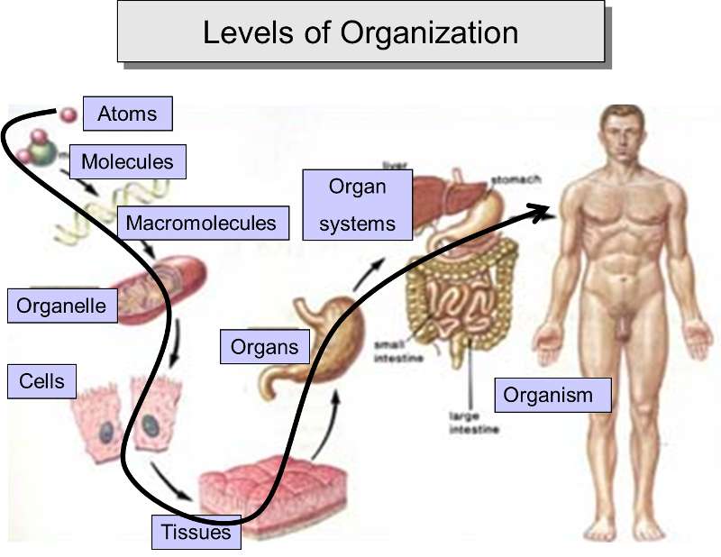
One can see that cells, the smallest unit that meets the criteria for being alive, are highly complex. Nevertheless, this complexity results from simple molecules being linked together to form a myriad of increasingly diverse and complex structures, and these, in turn, provide the basis for an even higher level of organization and complexity by assembling into macromolecular complexes, such as membranes, organelles, microtubules, and lipoproteins. From the cellular level, one can then envision the aggregation of cells to form tissues, which become the basis for organs and even organ systems in an incredibly diverse array of multicellular organisms.

Adapted from: http://www.theorganicstartupbook.com/2012/07/07/evolutionary-levels-sublevels-4-of-5/


