Dermal Monitoring
Skin is considered to be the largest organ and serves many functions. On the one hand, skin serves as a protective barrier from the external environment (physical, chemical, and biological barrier). Skin also plays an important role in absorption by allowing some substances to pass through the skin and into the bloodstream. While some substances such as vitamins from the sun play an important role in maintaining health, harmful substances can also be absorbed through skin.
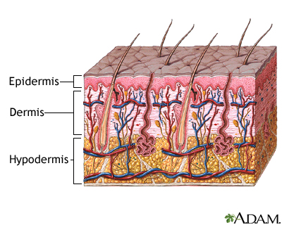
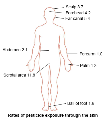
As shown in the image below on the left, there are three main layers that make up the skin: the epidermis, the dermis, and the hypodermis. The stratum corneum is the outermost portion of the epidermis and consists of dead cells, which are replaced every 2-3 weeks. The make up of this layer is such that it functions to protect underlying tissues from dehydration as well as chemicals and mechanical stress. It acts as a rate-limiting diffusion barrier. The image on the right highlights how different parts of the body can absorb chemicals such as pesticides through the skin at different rates. Note the areas that have the highest rate compared to the lowest rate.
Skin Permeability
Understanding the dynamics of how chemicals can be absorbed through the skin reveals some important factors that influence skin absorption. Chemicals penetrate the skin through passive diffusion, meaning a transfer of a given chemical through the skin occurs when there is a concentration gradients. Steady-state rate of transfer across a homogeneous membrane is determined by Fick's first law of diffusion shown in the table below.
Fick's First Law of Diffusion
- Jss= steady-state flux or rate of mass transfer (mg/cm2-h)
- Kmv = partition coefficient accounts for equilibrium differences between vehicle and the membrane (dimensionless)
- D = diffusivity of substance within membrane (cm2/hr)
- L = thickness of membrane (cm)
- C = concentration gradient (mg/cm3)
Measuring Dermal Exposure
Experimental results are frequently summarized as a permeability coefficient Kp (cm/hr), which is used to calculate absorbed dermal dose rates in a given scenario. Kp values are chemical-specific and are essential for characterizing transport through skin and are often calculated as a function of Kow and molecular weight. The EPA uses the following formula to calculate the Kp.
Skin Permeability Coefficient (Kp)
- Kp = permeability coefficient (cm/h)
- Kow = octanol/water partition coefficient
- MW = molecular weight (g/mol)
** It's important to note that the relationship expressed in this formula does not hold well for small polar molecular weight compounds or for large lipophilic compounds.
There are three general categories of dermal exposure assessment methods highlighted below. All of the following methods measure mass of material on the skin surface, though each measures slightly different aspects of dermal exposure.
Fluorescent tracer techniques
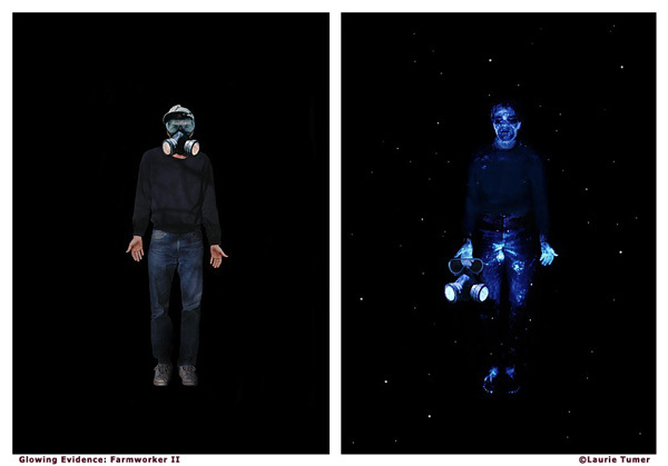
Fluorescent tracer techniques rely on the measurement of UV fluorescence from materials deposited and retained on the skin. This method is often used to mimic pesticide exposure in which a fluorescent tracer is mixed, diluted, and applied like pesticides. Then a blacklight is used to reveal the patterns and areas of exposure to the skin. The image below highlights photography from Laurie Tumer, an environmental artist, in which this method is performed and the areas of exposure are revealed on the subject.
Removal techniques
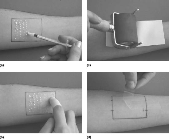
Removal techniques where substances deposited on the skin are removed generally by rinsing, wiping, or tape stripping. The image below provides an overview of the tape stripping procedure. In this case, a substance is first applied to the skin (a & b) which do not always apply since investigators are often looking for environmental exposures, which does not require applying the agent. The last two images are the key steps in which a strip of adhesive tape is applied to an area of skin following exposure (c) then the tape is removed with the agent of interest (d) as well as several micrometers of the stratum corneum.
Surrogate skin techniques
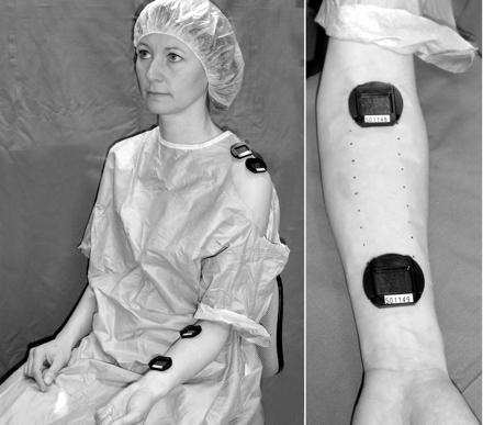
Surrogate skin techniques rely on a collection medium placed against the subject's skin, such as patches or gloves. The image below provides an example of using patches both on the shoulder and forearm, which are used as collection media.
Practical Application
Serum PBDEs in a North Carolina Toddler Cohort: Associations with Handwipes, House Dust, and Socioeconomic Variables is an article by Stapleton et al. The article looks to characterize dermal exposure to PBDEs in household dust. More specifically, investigators wanted to examine the relationship between household dust, dermal exposure, and serum levels of PBDEs. Review the introduction and study methods prior to answering the questions below the article.

