


Cardiovascular System
The primary function of the heart and blood vessels is to transport oxygen, nutrients, and byproducts of metabolism. Oxygenated and nutrient rich blood is distributed to tissues via the arterial system, which branches into smaller and smaller blood vessels from arteries to arterioles to capillaries (where most exchange occurs). Deoxygenated blood and metabolic byproducts are returned from capillaries via venules and then vein. The heart functions as a pump to maintain circulation. The heart is a discrete organ, which in humans has four distinct chambers. Conceptually, there is the right side of the heart (right atrium and right ventricle) which receive blood returning from the periphery and send it to the lungs (via the pulmonary artery) for re-oxygenation. Once blood is re-oxygenated in the lungs it is returned to the left side of the heart via the pulmonary veins. After entering the left atrium, blood enters the left ventricle and is pumped into the aortic arch for distribution to the entire body. The illustration below provides a schematic representation.
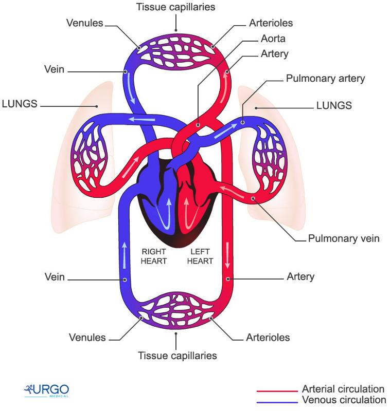
Source: http://humananatomybody.info/neck-arteries-model-labeled/
Note also the the heart is just like any other tissue in that it needs a continuous supply of oxygen and nutrients. The receives its blood supply from coronary arteries, which arise from the root of the aorta. The right and left coronary arteries are shown in the illustration below.
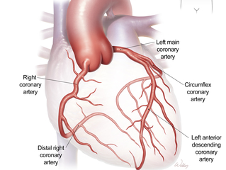
Source: https://www.bcm.edu/healthcare/care-centers/cardiothoracic/procedures/coronary-artery-disease-coronary-bypass
Normal Structure of Blood Vessels
All blood vessels (arteries and veins) have three primary layers: the intima, media, and adventitia. Normally, the walls of an artery are smooth, allowing unobstructed blood flow. The innermost layer of a blood vessel (the intima) is lined with endothelial cells, which are in direct contact with blood. This is shown at the top of the schematic cut-away diagram below.
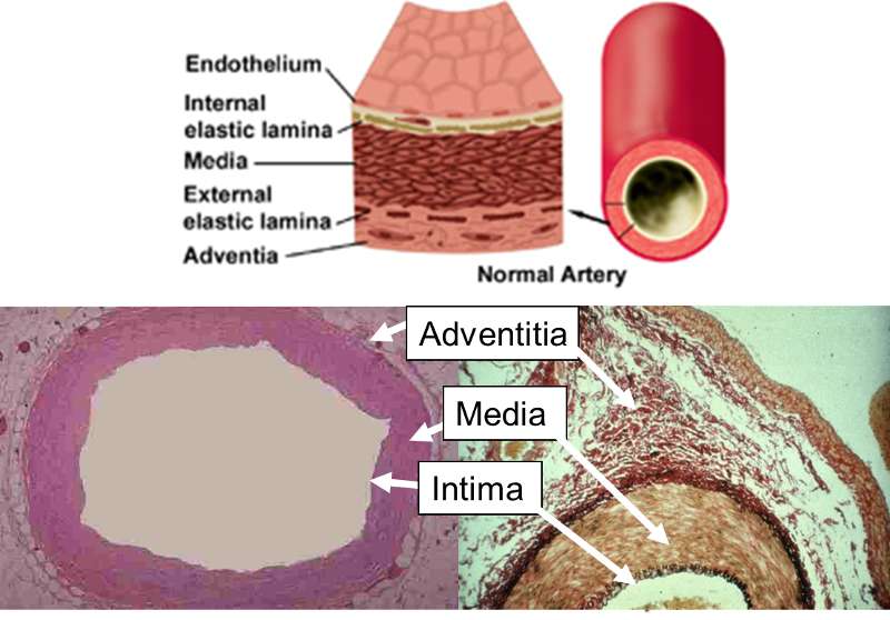
The internal elastic lamina is the barrier between the intima and the underlying media or "tunica media." The media media consists of multiple layers of smooth muscle cells which control the diameter of the blood vessel by contracting or relaxing in response to neural and chemical signals. The outermost layer is the adventia, which consists of connective tissue and also contains nerves and small blood vessels supplying the artery itself.
The next image is a scanning electron micrograph of an arteriole showing the layers of the artery and blood cells criculation within the lumen adjacent to the endothelium.
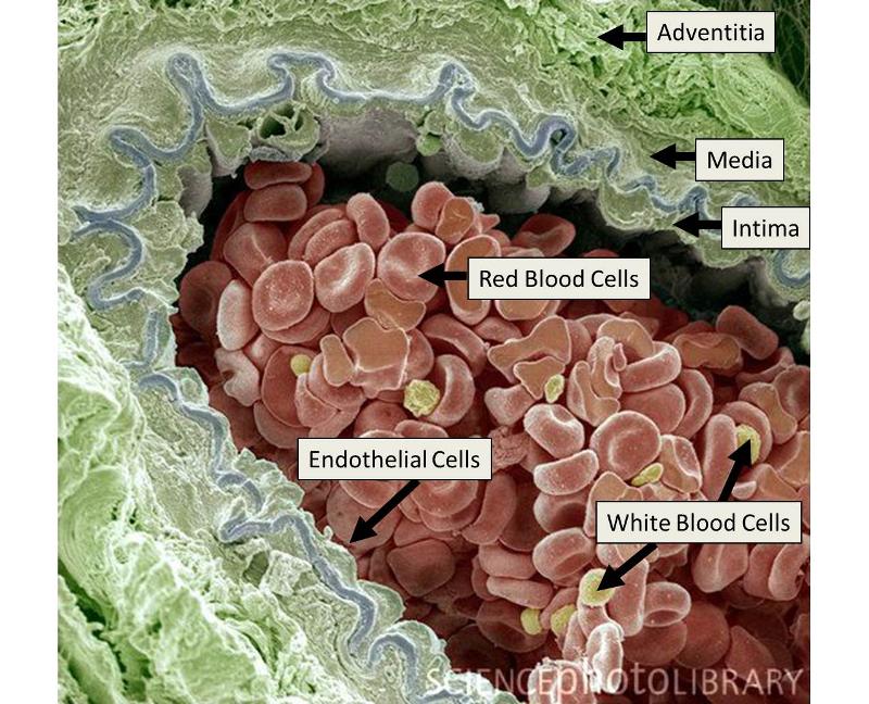
Source: Illustration adapted from: http://www.divingfollonica.com/large-elastic-arteries&page=5
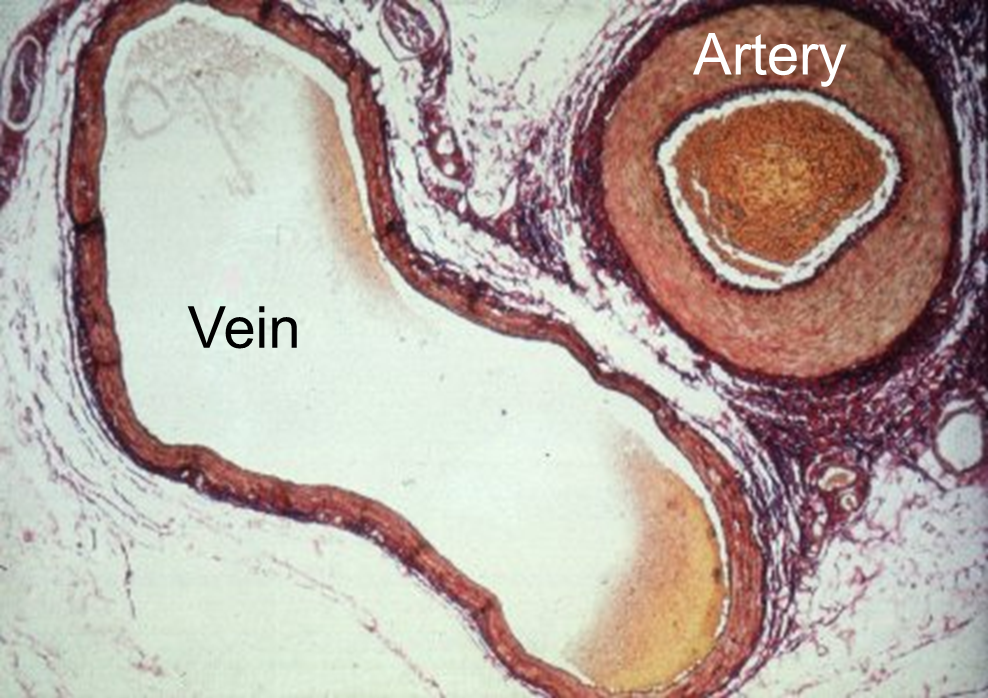
This three layered structure is characteristic of both veins and arteries, but veins have thinner walls, because the media is less developed. The image below provides a comparison of a vein and an artery.
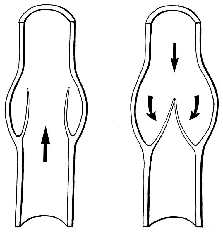
Another difference between arteries and veins is that veins can have internal valves, which help in maintain blood flow back to the heart. Without valves in the veins of the leg, venous blood would tend to pool in the lower leg when standing or sitting. The uni-directional valves (see the drawing on the right) prevent blood from flowing back down.