Infectious Agents
Source:http://www.ask.com/health/word-pathogen-mean-cdf78af549ca891e
This module will provide an overview of infectious agents, with an emphasis on the agents that are currently most relevant.
After successfully completing this section, the student will be able to:
 and give examples of each.
 epidemic in cattle caused disease in humans.

Biologists generally classify living organisms into one of the five kingdoms illustrated here. Bacteria are the most primitive and likely represent the earliest living organisms, from which the protista and other kingdoms are likely to have evolved. The overlap of the kingdoms in this figure is intentional, because, given the evolution of increasingly complex and diverse species, there are no clear-cut dividing lines, and classification has sometimes been ambiguous. Fungi, for example, were once classified with plants, but some of their structural characteristics are quite distinct from those of plants, and they are now classified in their own kingdom.
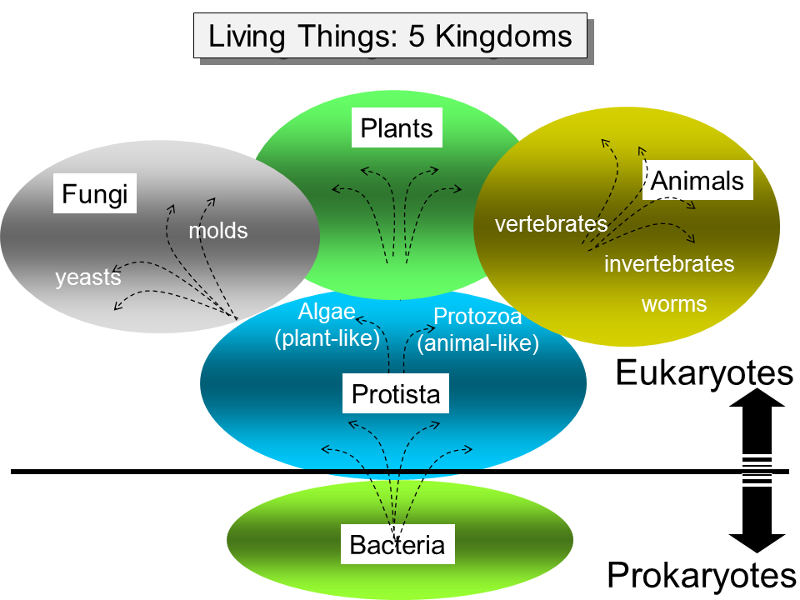
Species from all five of the kingdoms have the potential to influence human health, either positively or negatively.
While natural selection implies competition among and within species, we are increasingly aware that there is a strong interdependence among species. For example, most bacteria are non-pathogenic and live on the inner and outer surfaces of our bodies in staggeringly large numbers. These "normal flora" actually outnumber the cells in our body, and they provide many benefits. A key benefit is that by living on our skin and on the epithelial lining of our respiratory, digestive, and uro-genital tract, these usually harmless bacteria prevent pathogenic species from gaining a foothold.
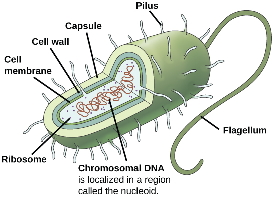
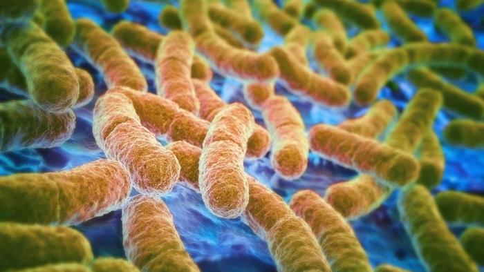
The bacteria are the oldest and simplest living organisms, and all of the bacteria are "prokaryotes," meaning that they do not have a true membrane-bound nucleus as eukaryotes do. [Prokaryote is derived from Greek,meaning "before nucleus"; eukaryote means "true nucleus."]
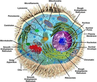
The figures and tables below provide a comparison of prokaryotic versus eukaryotic cells.
|
Prokaryote
Source: https://www.boundless.com/biology/textbooks/boundless-biology-textbook/prokaryotes-bacteria-and-archaea-22/structure-of-prokaryotes-141/basic-structures-of-prokaryotic-cells-562-11775/ |
Eukaryote
Source: http://dinopedia.wikia.com/wiki/Eukaryote |
|
Prokaryotes |
Eukaryotes |
|---|---|
|
Generally smaller (0.2-2.0 μm) |
Generally 10-100 μm |
|
No nuclear membrane. There is generally a single circular chromosome composed of DNA |
Have a true nucleus, consisting of nuclear membrane & nucleoli. Eukaryotes have multiple linear chromosomes. |
|
No membrane-enclosed organelles. |
Membrane-enclosed organelles include lysosomes, Golgi complex, endoplasmic reticulum, mitochondria & chloroplasts |
|
Flagella are simple, consisting of just two protein building blocks |
Flagella are complex and are composed of multiple microtubules |
|
Present as a capsule or slime layer |
Present in some cells that lack a cell wall |
|
Frequently have a cell wall and a cell cell membrane. The cell membrane lacks carbohydrates and generally lacks sterols |
Usually do not have a cell wall. The cell membrane does have sterols and carbohydrates that serve as receptors. |
|
Lack a cytosketeton |
Have a cytoskeleton and can perform cytoplasmic streaming |
|
Binary fission |
Mitosis |


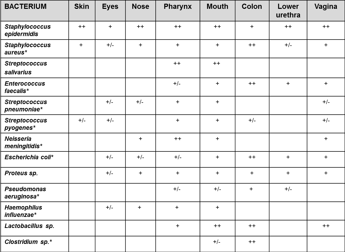
We are learning more and more about the benefits of bacteria. Certainly, with the help of fungi, bacteria play a vital role in breaking down and recycling dead organisms. Healthy internal tissues (e.g. blood, brain, muscle, etc.) are free of microorganisms, but skin & mucous membranes in our gastrointestinal tract, our respiratory tract, and our genito-urinary tract, are in contact with organisms in the environment, and these surfaces become colonized with many of these bacterial species. These bacteria that are regularly found at a given site are referred to as "normal flora." The normal flora of humans is consists of >200 species of bacteria. Their makeup depends on age, sex, stress, nutrition, etc. The table below shows a partial list of some of the more common bacteria regularly found in and on humans. The number of plus signs indicates their relative abundance. +/- indicates that the species may or may not be present.
Source: http://www.google.com/patents/WO2008049231A1?cl=en
These normal flora provide us with many benefits, which include:
Some data suggests that inappropriate use of antibiotics and the avoidance of microbes through disinfecting ourselves and our environment may have adverse effects on health. In fact, there is data to suggest that over-disinfection in children may increase their risk of autoimmune disease, obesity, and asthma. Here are several interesting links that provide some insights into this idea.
NPR Blog
This site provides an excellent perspective on the many benefits of bacteria. There is an 8 minute audio file that was aired on NPR, and there are many other interesting links.
http://www.npr.org/blogs/health/2013/07/22/203659797/staying-healthy-may-mean-learning-to-love-our-microbiomesGut Bacteria May Affect Our Brain
Lewis Thomas
An excerpt from Lewis Thomas: "The Lives of a Cell - Notes of a Biology Watcher." Bantam Books, 1974. pp.88-89."Germs"
Babies Know: A Little Dirt Is Good for You
From The New York Times: Personal Health: by Jane E. Brody, Published: January 26, 2009
Jonathan Eisen: Meet Your Microbes
This is a TED talk in which microbiologist Jonathan Eisen discusses the benefits of microbes and presents some ideas about how we might use microbes to improve human health. (14:23)
The Ecology of Disease
The Ecology of Disease is is an article by Jim Robbins from the New York Times. Robbins says:
"If we fail to understand and take care of the natural world, it can cause a breakdown of these systems and come back to haunt us in ways we know little about. A critical example is a developing model of infectious disease that shows that most epidemics — AIDS, Ebola, West Nile, SARS, Lyme disease and hundreds more that have occurred over the last several decades — don't just happen. They are a result of things people do to nature.
Disease, it turns out, is largely an environmental issue. Sixty percent of emerging infectious diseases that affect humans are zoonotic — they originate in animals. And more than two-thirds of those originate in wildlife."
Dirt in Our Diets
This is an OpEd piece from the New York Times on June 20, 2012.
Feces Infusion to Cure Overinfection With C. difficile
(report in New England Journal of Medicine, Jan. 16, 2013
While only about 5% of bacterial species are pathogenic, bacteria have historically been the cause of a disproportionate amount of human disease and death. There is good evidence that from the 1300s through the 1800s tuberculosis, bacterial pneumonia, typhus, plague, diphtheria, typhoid, cholera, dysentery were major causes of disease and premature death in Europe and the United States. Among those born in the United Kingdom in the 1800s, it is estimated that 70% died before the age of 25, and a large proportion of these deaths were due to bacterial infections. Not surprisingly, this burden of disease and early death fell most heavily on the poor. During the 19th century, however, there was the emergence of "the sanitary idea" in England and the United States, and the efforts to provide better waste disposal, clean water, better nutrition, and better working conditions were rewarded with remarkable reductions in disease and death rates.
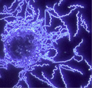
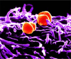
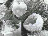
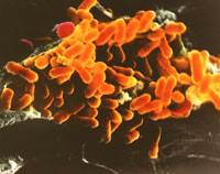
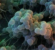
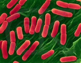
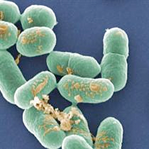
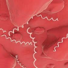
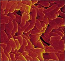
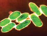
Nevertheless, bacterial pathogens still pose a threat. The table below provides microscopic images of a variety of bacterial pathogens that are relevant to disease in humans. The hyperlinks provide additional information, primarily from CDC summaries.
|
Borrelia burgdorferi (Lyme disease) |
Neisseria gonorrhea |
|
Chlamydia |
Rickettsia |
|
Cholera |
Salmonella |
|
Clostridium (tetanus, botulism, gas gangrene) |
Staphylococcus (Food poisoning, MRSA) |
|
E. coli |
Streptococcus |
|
Listeria |
Treponema pallidum (syphilis) |
|
Mycobacterium tuberculosis (TB) |
Yersinia pestis (bubonic plague) |
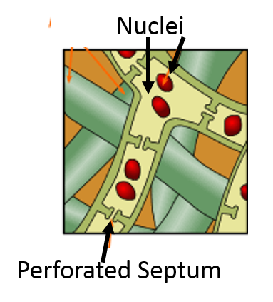
Fungi are plant-like and were once classified with plants, but fungi lack chlorophyll and differ in other ways from plants, so they are now classified in a separate kingdom. However, they are often mutualistic, living with other species, e.g. lichens (algae + fungus), and growing in a symbiotic relationship on rocks & trees. Fungi are saprophytes that decompose dead organic matter by growing into a substrate & absorb nutrients from it. Parasitic fungi often feed on living organisms without killing them (e.g. ringworm & athletes foot). Fungi are structurally different from plants. Fungi are generally composed of branching filaments ("hyphae") that sometimes form a large interlacing mass called a "mycelium." The hyphae have cross walls, but they are perforated allowing free passage of nuclei and cytoplasm. Unlike plants, fungi are not truly multi-cellular.
Like bacteria, fungi provide many benefits. In conjunction with bacteria, they are important in breaking down dead organic material & recycling nutrients through ecosystems. Fungi supply nutrients to the roots of many plants, and they also provide a source of antibiotics. In addition, they provide food (mushrooms, truffles), make bread rise, and enable fermentation of sugar to alcohol. The tabbed activity below shows some of the forms the fungi take that may be familiar to you.
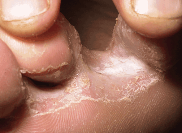
Like bacteria, fungi can comprise part of our normal flora. For example, Candida species are common fungi that are normal flora on our skin and in our respiratory, genital, and digestive tracts. Candida is normally kept in check by other normal flora as well as our immune system. Yet they can cause infections which can be mild (diaper rash and vulvar rash) or severe (oral thrush, esophagitis, and rapidly fatal systemic diseases in immunocompromised people). Candida is usually a yeast, but can form hyphae.
Fungi also cause a number of diseases in plants and animal e.g. ringworm, athlete's foot. Because fungi are more chemically and genetically similar to animals than to other organisms, fungal diseases are often very difficult to treat.
From the CDC web site:
"Fungi interact with humans, animals, and plants in a variety of ways. Some of these interactions can be beneficial; for example, both penicillin and bread use ingredients made from fungi. However, certain types of fungi can be harmful to health. Like bacteria and viruses, some fungi can act as pathogens. Human fungal diseases can occur due to infection or fungal toxins.
Why are fungal diseases a public health issue?
Mycotic (fungal) infections pose an increasing threat to public health for several reasons. The scientific and medical staff of the Mycotic Diseases Branch is involved with prevention and control among three broad categories of fungal infections:
The table below provides some examples of fungal diseases, with links to additional information sheets from the CDC.
|
Candida albicans |
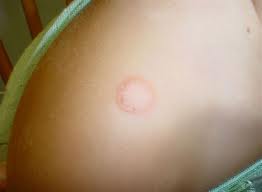
Dermatophytes (e.g., ringworm) |
|
Cryptococcus neoformans |
Dermatophytes (e.g., ringworm) |
Fungi also produce some interesting toxins which are products of a fungus' secondary metabolism, i.e. that part of fungal metabolism that is not essential for cell growth and maintenance of basic cell function. Why fungi produce such substances is not entirely clear, but they may, at least in part, be used for "chemical warfare" and provide some survival advantage in the environment.
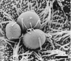
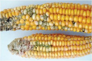
Aspergillus mold can grow on peanuts, corn, and grain.
Ergot fungus infects various cereal plants and forms compact black masses of branching filaments that replace many of the grains of the host plant.

Aspergillus mold is virtually unavoidable. Outdoors, it's found in decaying leaves and compost and on plants, trees and grain crops. The spores thrive in air conditioning and heating ducts, insulation, and some foods and spices. Aspergillus is so common in old buildings, even in older hospitals, that small epidemics have occurred among people with weakened immune systems when nearby buildings have been torn down. Everyday exposure to aspergillus is rarely a problem for people with healthy immune systems.
Certain species of the Aspergillus mold can produce toxins (aflatoxins) that can be metabolized to chemicals that have a high affinity for DNA. Their binding to DNA can cause mutations that can contribute to the development of a cancer. Aflatoxins are carcinogenic in rats, ducks, mice, trout & subhuman primates. Trout are the most susceptible; just 1 ppb (part per billion) of aflatoxin B1 will cause liver cancer in trout. Some studies indicate that aflatoxins increase the risk of liver cancer in humans who are hepatitis B carriers.
In 2004 there was a widespread outbreak of human aflatoxin poisoning in eastern and central Kenya as a result of aflatoxin contamination of locally grown maize, which occurred during storage of the maize under damp conditions. Affected individuals had jaundice and a high case-fatality rate.
Ergotism is a condition caused by poisoning with an alkaloids produced by the Claviseps pupurea fungus which infects cereals, especially rye. Intoxication can produce neurological symptoms (e.g., a burning or prickling sensation, or hallucinations, or psychosis). Some believe that ergot poisoning was responsible for the strange behaviors observed among young women in Salem, which resulted in the Salem witch trials. Ergotism can also cause severe constriction of blood vessels, resulting in gangrene. Episodes of ergotism occurred periodically in the Middle Ages, when it was known as "St. Anthony's Fire" because of the sensation of burning skin which occurred with the gangrenous form. Developed countries have controlled ergotism through carefully monitoring, but it still remains a potential threat in developing countries.
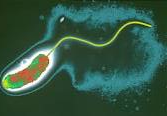
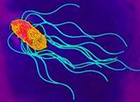
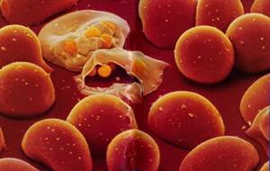
Protozoa are single-celled eukaryotes that ingest food (algae and bacteria) by phagocytosis and generally move via pseudopods (flowing extensions of the plasma membrane) or whip-like flagella. Most are too small to be seen with the naked eye, but can easily be found under a microscope. Protozoa reproduce by fission. The short video below provides more information and an illustration of protozoa.
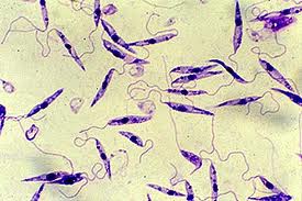
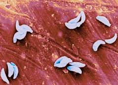
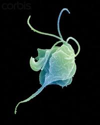
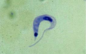
There are a number of protozoa that inhabit the gastrointestinal tract of humans. Most of these are harmless or cause only mild problems, but others cause serious disease. Many of the protozoa that infect humans are transmitted via the fecal-oral route, but others are transmitted via insect vectors (e.g., malaria and leishmaniasis), and trichomonas vaginalis is a sexually transmitted disease.
Some have fairly complex life cycles that may include a cyst stage that enable the organism to remain dormant in the environment for a period of time until a new host is acquired (e.g., malaria). Four life cycles are shown below.
|
Cryptosporidium Life Cycle |
Giardia Life Cycle |
|
Leishmaniasis Life Cycle |
Malaria Life Cycle |
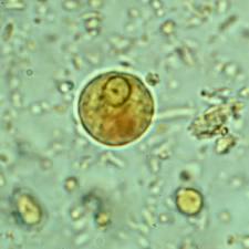
The table below provides illustrations and links to additional information for a variety of animal-like protozoa.
|
Cryptosporidiosis |
Entameba histolytica |
|
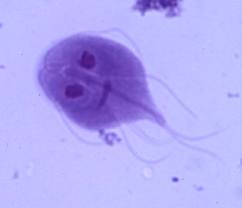
Giardia lamblia |
Leishmaniasis |
|
Toxoplasmosis |
Malaria |
|
Trichomonas vaginalis |
Typanosomiasis (sleeping sickness) |
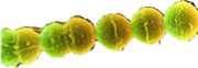
There are also plant-like protista (algae) that are responsible for producing so-called "red tides." These are caused by organisms called dinoflagellates that can produce neurotoxins. Fish or mollusks that eat these dinoflagellates concentrate the toxins, and, if humans eat affected fish or mollusks, severe illness or death can result. Actually, the term "algae" is used somewhat loosely to describe a rather broad group of organisms, which includes some species that are regarded as plants by some biologists and other species that are regarded as protista. For a more complete discussion of this confusing area, see "What are algae?".
Toxic blooms can also be caused by certain bacteria, such as the Microcystis that from Lake Erie that poisoned Toledo, Ohio's water supply in August 2014. The frame below provides access to a National Geographic article on the problem in Toledo.
From the CDC web site:
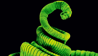
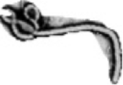
"Helminths are large, multicellular organisms that are generally visible to the naked eye in their adult stages. Like protozoa, helminths can be either free-living or parasitic in nature. In their adult form, helminths cannot multiply in humans. There are three main groups of helminths (derived from the Greek word for worms) that are human parasites:
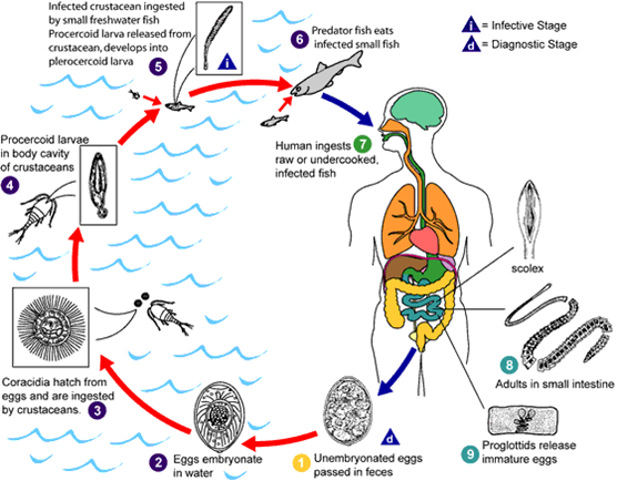
These organisms have fairly complex anatomy and they tend to have complex reproductive cycles as well. Note, however, that in order for humans to become infected with the fish tapeworm, the feces of infected human needs to find its way into water, and humans have to eat raw fish from contaminated waters.
|
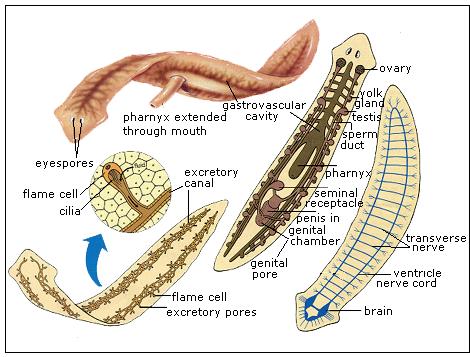
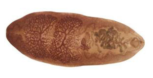
Examples of Flatworm Anatomy and Life Cycle |
|
|---|---|
|
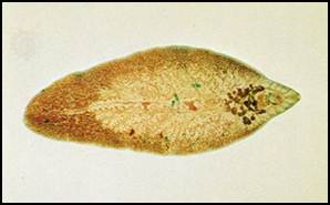
Liver fluke anatomy (a flatworm) |
Life Cycle of the Fish Tapeworm |
|
|
|
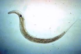
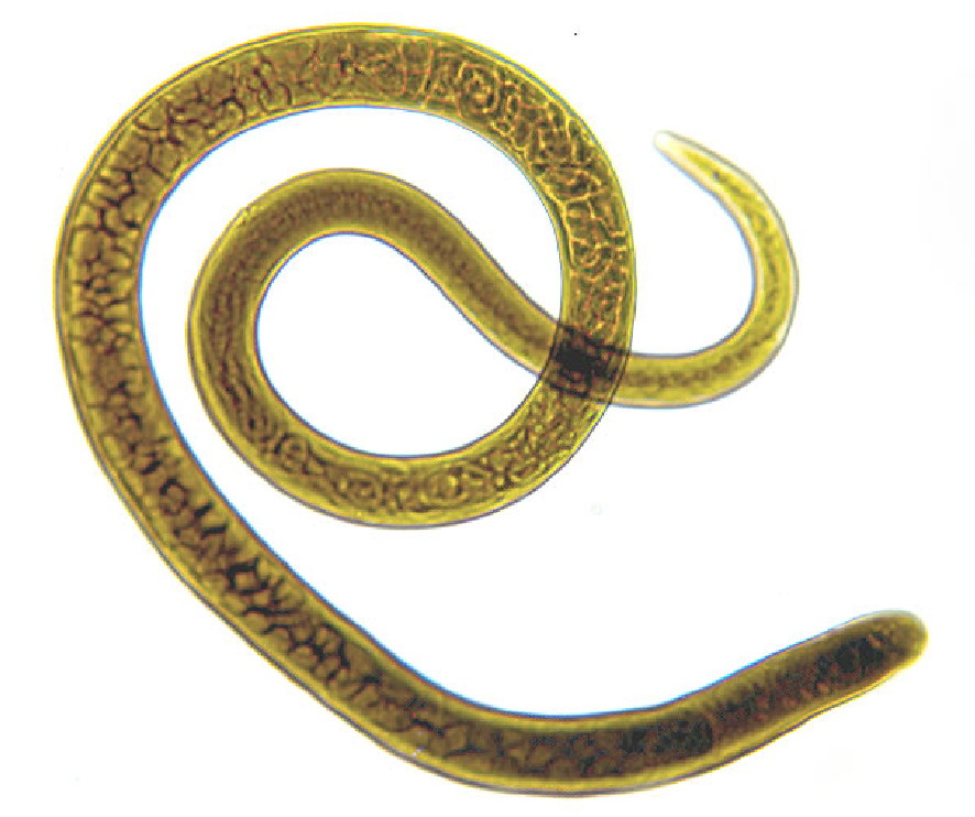
The next table (below) illustrates the anatomy and life cycle of a hookworm, a roundworm.
|
Hookworm (a round worm) Anatomy and Life Cycle |
|
|
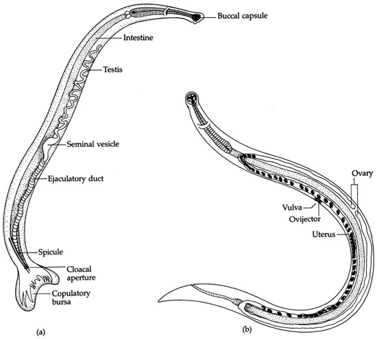
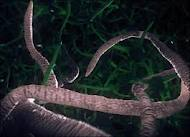
Adult Male (left) and Female (right) Hookworms |
|
|
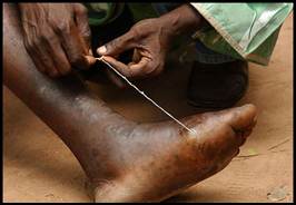
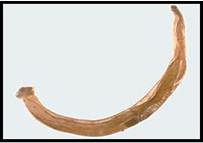
Guinea worm See life cycle to the right. |
|
See the links below to learn more about the campaign to eradicate Guinea worm. The first is a link to a short article in The Washington Post: "Guinea Worm is poised to become the second human disease to be eradicated" - published August 27, 2012. The next two links describe simple water filtration devices that have played a major role in the eradication campaign.
Study the life cycle of the guinea worm in the table above; under what circumstances is it potentially possible to eradicate an infectious disease of humans?
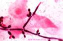
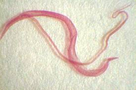
The table below provides a 'rogues gallery' of flatworms and roundworms that can infect humans.
|
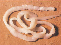
Flatworms (Platyhelminths) |
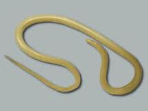
Roundworms (Nematodes) |
|---|---|
|
Intestinal fluke |
Ascaris |
|
Liver fluke |
Filariasis |
|
|
|
|
Fish tapeworm |
Hookworm |
|
Beef tapeworm |
Pinworm |
|
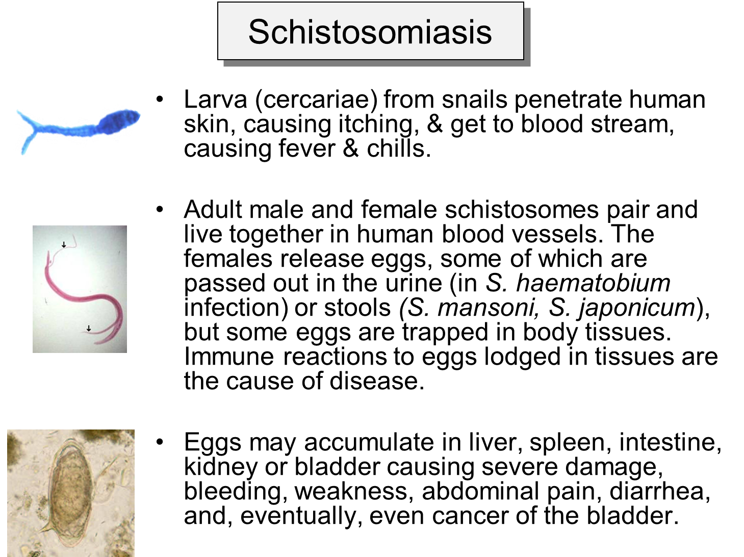
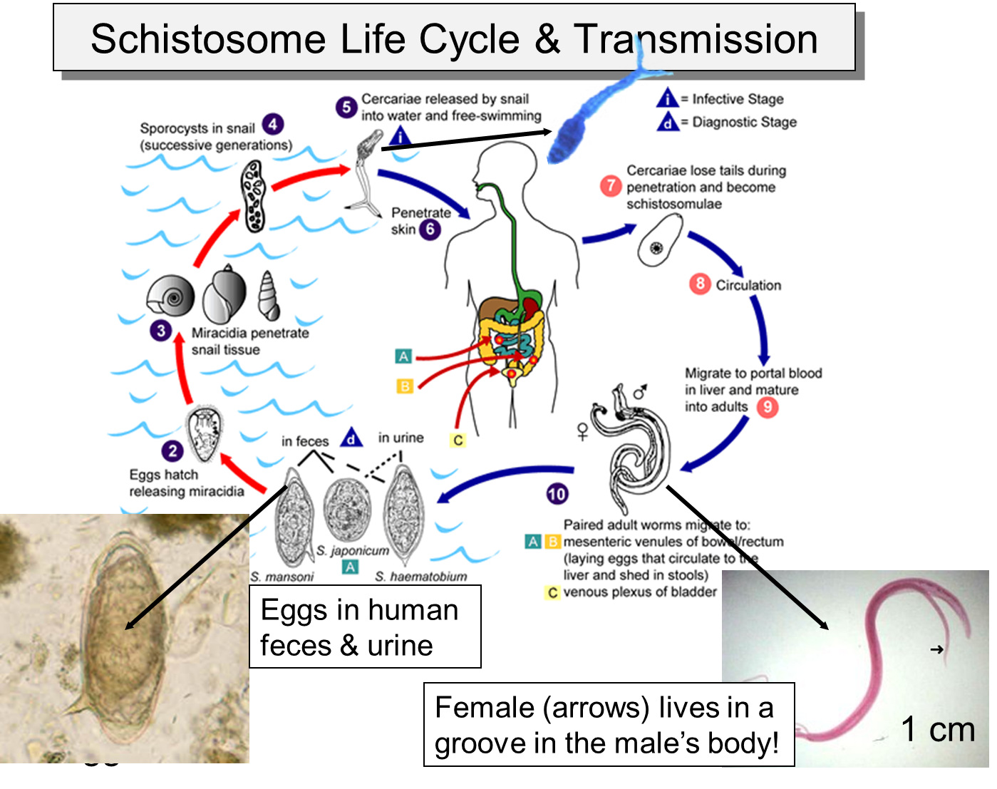
Schistomsomiasis |
Trichinella spiralis (Trichinosis) |
From the CDC web site:
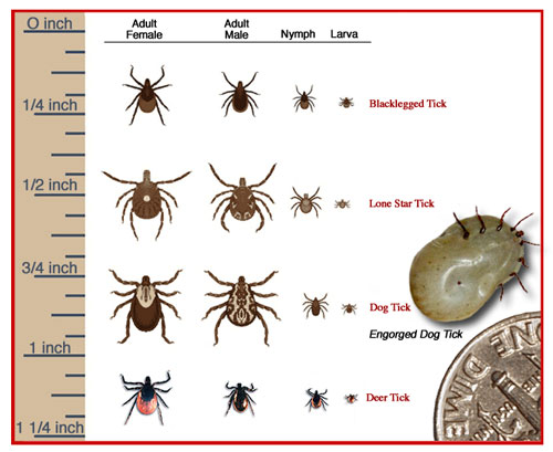
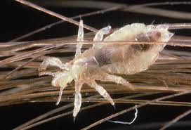
"Although the term ectoparasites can broadly include blood-sucking arthropods such as mosquitoes (because they are dependent on a blood meal from a human host for their survival), this term is generally used more narrowly to refer to organisms such as ticks, fleas, lice, and mites that attach or burrow into the skin and remain there for relatively long periods of time (e.g., weeks to months). Arthropods are important in causing diseases in their own right, but are even more important as vectors, or transmitters, of many different pathogens that in turn cause tremendous morbidity and mortality from the diseases they cause."
|
Ticks |
A louse |
This link is to an online learning module on Lyme disease.
Scientists generally agree that there are four requisite characteristics of living organisms.
All of the organisms belonging to the five kingdoms of living things in the previous sections of this module have all five of these characteristics. However, there are two types of infectious agents that do no meet these criteria:
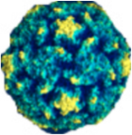
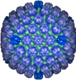
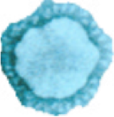
Viruses are assembles of organic molecules that consist of some short strands of RNA or DNA encapsulated within a protein shell. They are often referred to as if they were living organisms, but they don't meet the criteria listed above for living things. In a sense, they perhaps represent a primitive assembly of organic molecules that resemble living cells, yet they do not have the complexity and the the characteristics needed to be truly living organisms that are capable of reproducing independently, responding to the environment, and capturing energy on their own. Instead, all of the viruses are all parasitic, because they all need a living host cell in order to replicate. Once they bind to living cells and get taken up, they can use a host's cellular energy and machinery (e.g., ribosomes) to replicate its genetic material and its proteins, and these can self-assemble into new virus particles. These can lie dormant, or they can cause the host cell to rupture, releasing the progeny virus particle, which can go on to infect other host cells. Viruses can infect all kinds of living cells, including bacteria, and almost all viruses are pathogenic. When viruses infect a host cell, they can cause disease through several possible mechanisms:
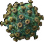
In the general process of infection and replication by a DNA virus (e.g., Herpes simplex, type 1, Herpes simplex, type 2, Varicella-zoster virus, & Human papillomavirus) a viral particle first attaches to a specific host cell via protein receptors on its outer envelope, or capsid. The viral genome is then inserted into the host cell, where it uses host cell enzymes to replicate its DNA, transcribe the DNA to make messenger RNA, and translate the messenger RNA into viral proteins. The replicated DNA and viral proteins are then assembled into complete viral particles, and the new viruses are released from the host cell. In some cases, virus-derived enzymes destroy the host cell membranes, killing the cell and releasing the new virus particles. In other cases, new virus particles exit the cell by a budding process, weakening but not destroying the cell.
Other viruses contain RNA in their core. The diagram below illustrates the events that take place during influenza infection.
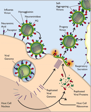
First, the virus attaches to a host cell (e.g., an epithelial cell lining the nasopharynx). The hemaglutinin protein (shown in green) on the exterior of the viral particle has a shape that enables it to bind to a neuraminic acid protein on the host cell. Binding triggers changes in the host cell membrane that cause it to invaginate and internalize the virus. The virus then sheds its protein coat, releasing its RNA into the cell. The viral RNA is used as messenger RNA to produce viral proteins. In this process the host cells ribosomes, amino acids, and ATP are used to create new viral proteins. One of the viral proteins that is replicated in a viral RNA polymerase that copies the viral RNA template to produce new copies of viral RNA for the replicated viral particles. Once there is a critical mass of new viral proteins and RNA, they self-assemble to form new viral particles that are released from the host cell either by budding off or by rupturing the cell membrane. These new viral particles can then infect additional host cells.
Still other RNA viruses, called retroviruses (e.g. HIV), use an enzyme called reverse transcriptase to copy the RNA genome into DNA. This DNA then integrates itself into the host cell genome. These viruses frequently exhibit long latent periods in which their genomes are faithfully copied and distributed to progeny cells each time the cell divides. The human immunodeficiency virus (HIV), which causes AIDS, is a familiar example of a retrovirus. The HIV life cycle is summarized in the illustration below.
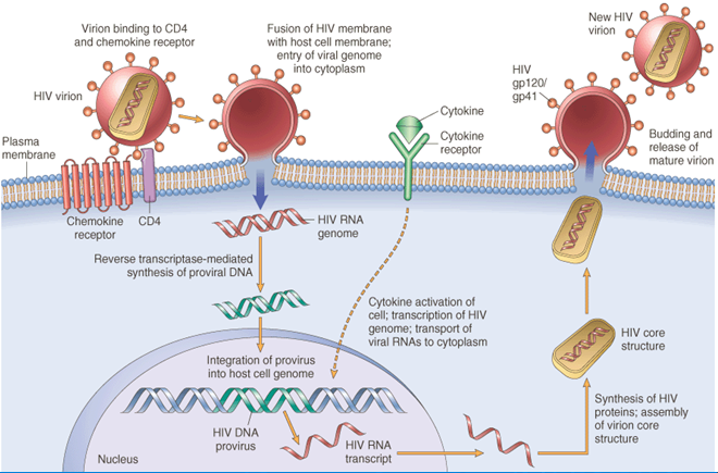
Source http://medicinembbs.blogspot.com/2011/02/diseases-of-immunity.html
This is a brief video that summarizes events that take place during HIV infection.
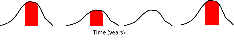
Viral infections can display several temporal patterns of disease. T


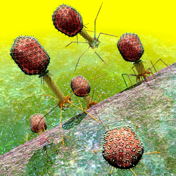
|
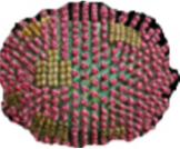
Bacteriophage |
Influenza Virus |
|
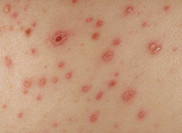
Chicken Pox (Varicella-Zoster) |
Mumps |
|
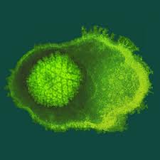
Ebola
|
Polio |
|
Herpes
|
SARS |
|
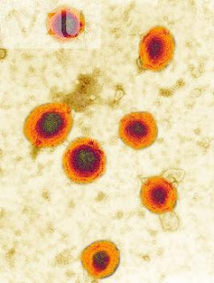
HIV |
Smallpox |
The term "prion" was coined from "protein infection particle." Prion diseases have been with us for some time, but only came to the attention of the general public during the epidemic of "Mad Cow Disease" that began in the United Kingdom in the 1970s. Prion proteins are normally found in all mammalian brains, but it is believed that altered forms of these proteins fold abnormally as a result of a mutations; as a result the proteins encoded by the mutant genes have distorted shapes, and they are not broken down normally.
Prusiner's Hypothesis:
(Dr. Stanley Prusiner was awarded a Nobel Prize in 1997 for his work on the discovery of prions.)
His hypothesis is that a mutant prion is an abnormally folded prion protein (PrPSc) that is resistant to heat & sterilization, and does not evoke an immune response. The protein is chemically the same, but folds differently. The abnormal protein contacts normal proteins in neural tissue and induces them to refold into an abnormal conformation as well. Refolded molecules induce the same change in still more proteins. The abnormal proteins resist degradation and accumulate in neural tissue causing damage. The illustration below shows an artists depiction of a normal prion protein (PrPC on the left) and an abnormal prion protein (PrPSc on the right). The abnormally folded protein has segments folded into "beta sheets" (the segments shown with thick green arrows). These segments tend to sticky together causing clumps of proteins that resist breakdown. Over time, the clumps grow larger and destroy nerve cells in areas of accumulation, leading to progressive neurological symptoms (so-called "prion diseases").
Source: http://www.scq.ubc.ca/prions-infectious-proteins-repsonsible-for-mad-cow-disease/
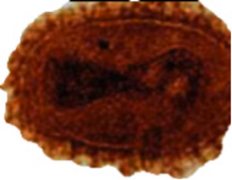
The deposition and accumulation of these abnormal proteins in the brain results in brain tissue that appears to be riddled with holes when the brain is sectioned and stained post-mortem. As a result, these disease are also referred to as transmisible "spongiform" encephalopathies. The image below shows a specimen of brain tissue viewed through a microscope. The light colored "holes" represent areas of the brain where deposits of the abnormal proteins destroyed cells.
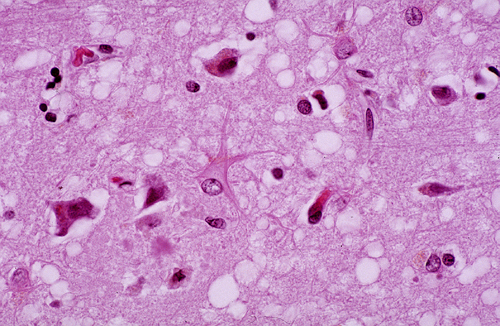
Source: http://neuropathology-web.org/chapter5/chapter5ePrions.html
The abnormal folding is believed to be initiated by a mutation. In the case of Creutzfeldt-Jakob Disease (CJD) there are two forms of the disease:
In addition, prion diseases are also transmissible if neural tissue with abnormal prion proteins is ingested or transmitted through medical procedures such as transplants or injections or via instruments contaminated with abnormal prion proteins. Kuru is a type of transmissible spongiform encephalopathy that occurred in New Guinea as a result of ritual cannibalistic practices that involved eating the brain tissue of human relatives who had died. At some point a mutation resulted in an abnormal prion protein that was transmitted among relatives as a result of eating affected brain tissue, which initiated a chain reaction in the the recipients that resulted in transmissible spongiform encephalopathy.
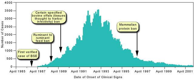
Scrapie is a disease of sheep that was described some time ago, and is now thought to be a prion disease. Many believe that the epidemic of bovine spongiform encephalopathy (BSE or "mad cow disease") that occurred in the United Kingdom in the 1970s was triggered when farmers began to feed cattle meat and bone meal that contained neural (nerve) tissue from scrapie-infected sheep or from cattle with spontaneously occurring BSE.
Source: http://neuropathology-web.org/chapter5/chapter5ePrions.html
The epidemic in cattle peaked in 1993 and is believe to have caused more than 184,500 cases of BSE. In conjunction with that epidemic a new human prion disease called variant Creutzfeldt-Jakob disease (vCJD) was first reported by the United Kingdom in 1996. There is evidence that vCJD resulted when humans ate meat or other animal tissues from cattle infected with BSE.
Link to CDC's web pages on prion diseases.